

Art Appreciation – July/August 2026
At the third attempt (although Molly insists it is the fifth) we had our much anticipated presentation on Anatomy and Art.
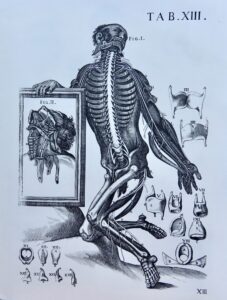 As an introduction, Molly shared some thoughts about why studying the human body, and getting beneath the surface to see what we are all made of, seems to have been something humans have done for millennia. She suggested that this arises, amongst other things, from our desire to explain, to record our knowledge, to create an image which might shock or even titillate, or produce an emotional response in the viewer. At some periods, it arises from exploring the new possibilities of technology as our ability to observe and record develops.
Some of the earliest explorations of the human body were undertaken by the ancient Egyptians as part of their mummification practices. Some of their inscriptions give detailed explanations of how they performed these rituals, showing the instruments they used and what they knew about anatomy.
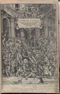
We know from their art that Japanese doctors dissected corpses many centuries ago, as did Greeks and Romans. Their ideas were followed slavishly well into the middle ages in Europe. The knowledge and ideas of the ancient world were revered until remarkably recently, and we were shown many images from as late as the 17th century of dissections being carried out just as Galen or Hippocrates advised. Typically, a corpse would have been opened up in front of an audience of students (all male, of course) with the learned teacher sat on a podium giving directions to the man with the scalpel. Not for nothing do we still use the term ‘operating theatre’, as these dissections were at least in part for spectators and attracted large audiences. Corpses were hard to obtain, usually sourced from executions or grave robbing, so a dissection was a major event. Molly showed us a number of images of dissections by artists which were primarily intended to demonstrate knowledge of human anatomy, but were often presented in dramatic poses – a fusion of art and science.
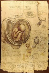 Many Renaissance artists were fascinated by the process of dissection and the new scientific knowledge. Leonardo da Vinci used this to create some of the most extraordinary images, including an unborn foetus. Our knowledge of how the foetus develops evolved over many years as it depended on the procurement of bodies of women at all the stages of pregnancy.
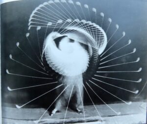
More recently, artists and scientists have been able to use new technologies such as photography and X-rays to extend their understanding of anatomy. We saw images produced by time lapse photography which showed for the first time in detail how the body moved during complicated actions such as swinging a golf club or jumping.
Some artists have also combined photography with sculpture to create some extraordinary and intriguing images. Microscopes have given artists another tool to explore our anatomy.
While many artists have focussed on portraying physical beauty, many modern artists such as Francis Bacon, are more interested in showing us how we really appear – with no compromise. Many of the pictures could not be described as beautiful but were nonetheless compelling.
The presentation raised questions about what constitutes ‘art’, the boundaries between art and science, and what art is for.
At our next meeting on Tuesday 11th August members are invited to select a work of art on the theme of ‘Journeys and Travel’.
Lynne Vick, Convenor